数字放射成像图像伪影
图1 shows a lateral chest image with an unusual superimposed pattern on the anatomy. This is an example of a CR image obtained with cassette reversed, where the tube side of cassette is pointed away from the x-ray tube source and toward the patient. Cassette plastic structural patterns are projected onto the imaging plate (particularly noticeable in the arms and anterior part of the patient. In this image a reversal of left to right can also unknowingly occur. 强烈建议使用铅标记.
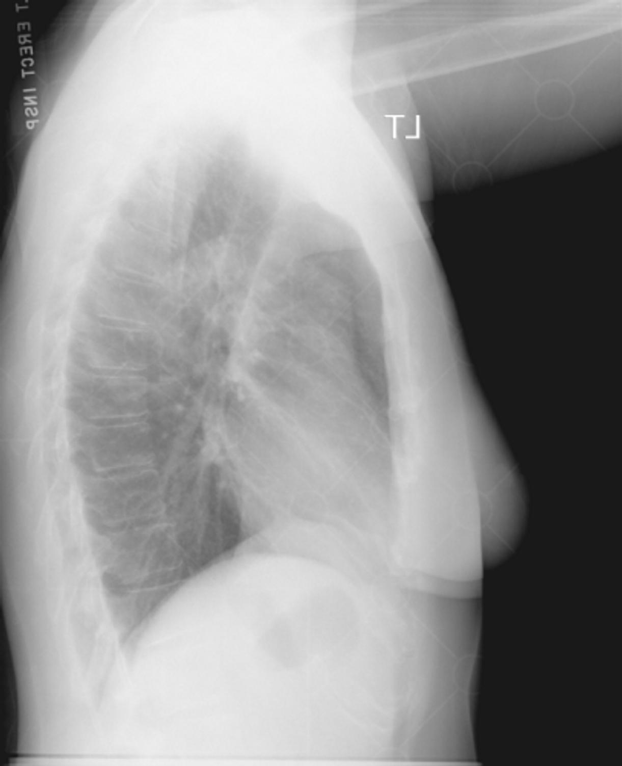 |
| 图1. Image patterns from the plastic support structures in the CR cassette are superimposed on the anatomy, caused by the placement of the imaging plate and the cassette upside down in the cassette holder.(点击图片查看完整版). |
垂直模式的高强度信号, 如图2所示, usually represent foreign materials that are stuck to the light path assembly that acquires the photostimulated luminescence signal from the CR imaging plate as it is being scanned by a laser beam. As the light is blocked at the same spot as the plate translated through the optical stage, 工件与激光束读数垂直, 在印版平移(慢扫描)方向. 条纹看起来很亮, since the image undergoes a reverse grayscale transformation to make the image appear similar to a screen-film image with processing.
 |
| 图2. Foreign materials on the light collection guide in a CR reader produces bright linear lines in the output image. (点击图片查看完整版). |
Digital detector system malfunctions can have a great impact on the quality of the output image. 在图3, an artifact caused by a galvanometer (the device that deflects the laser beam in a CR reader rapidly across the imaging plate as it translates through the optical stage) and digitizer becoming unsynchronized is shown. 图像列不能正确排列并移位.
 |
| 图3. A failure of the galvanometer and digitizer to synchronize during readout causes catastrophic image quality problems as shown. (点击图片查看完整版). |
Problems not directly associated with the digital detector system can also be manifested in the image. 图4 shows image artifacts caused by a metal filter in collimator that became unfastened and mis-positioned, projecting a variation in x-ray fluence across the anatomy and onto the detector. Similar artifacts are caused by CR imaging plates that are not erased frequently and/or exposed to x-ray scatter from another procedure, resulting in a variable background signal that is superimposed on the image. Double exposures are also common, since the imaging plate is a reusable detector.
 |
| 图4. 工件 in this image are caused by filters in the collimator assembly of the x-ray tube becoming loose and mispositioned in the x-ray beam, attenuating the x-ray beam non-uniformly and resulting in the attenuation pattern shown in the image. (点击图片查看完整版). |
工件 due to "aliasing" arise as a result of insufficient sampling of high frequency digital signals in an image represented by sharp edges or periodic structures such as anti-scatter grid lines. The high frequency signals masquerade as low frequency signals that are superimposed over the total image. Also known as moiré patterns, the information content of the image is compromised. A typical antiscatter grid used in an oscillating "Bucky" assembly has a grid frequency of 40-50 lead strips/cm, 哪个是大多数数字系统可以分辨的, but beyond the Nyquist frequency (highest frequency in the image containing "useful" resolution). 高频栅极的使用(例如.g., 70 grid strips/cm) will help eliminate aliased signals as those signals are beyond the resolvability of the digital detector system.
图5 illustrates how high frequency signals that are insufficiently sampled can result in lower frequency signals in the digitized output image.
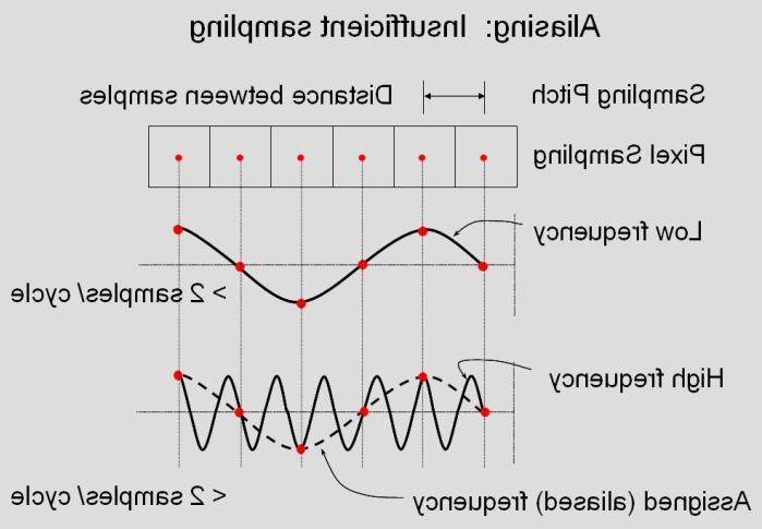 |
| 图5. 这张图说明了混叠是如何发生的. 在一个离散的数字检测器, the sampling pitch (space between samples) is determined by the detector dimensions. Each "sample" occurs in time at the center of the sampling aperture. 对于低频信号, having at least 2 samples / frequency cycle allows the computer system to correctly assign the given response of the varying signal; however, if the sampled signals contain frequencies that are sampled at less than 2 samples / cycle, then the computer system assigns a lower frequency signal than the actual frequency signal. The assigned frequency represents an aliased signal in the output image. |
Aliasing or moiré patterns can easily be caused by low frequency grids in a digital image, because of the very high contrast signal that the grid strips project onto the detector that are beyond the "Nyquist frequency" but still resolvable by the digital detector. 如图6所示.
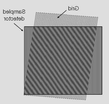 |
| 图6. This illustration demonstrates a low frequency beat signal generated by the superimposition of a grid structure on a sampled array. |
临床, 混叠信号可能相当突出和压倒性, as shown in the lateral skull image in 图7 acquired with a low frequency, 固定栅板.
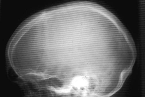 |
| 图7. Aliasing artifact is superimposed on a lateral skull image using a CR detector. (图片由北卡罗来纳大学巴里·伯恩斯提供). |